
Quick Summary: Image recognition for medical diagnosis uses artificial intelligence to analyze medical images like X-rays, MRIs, and CT scans to detect diseases, tumors, and abnormalities. Deep learning algorithms, particularly convolutional neural networks, can identify patterns in imaging data with accuracy comparable to or exceeding human radiologists in specific tasks. The FDA has cleared 1,451 of AI-enabled medical imaging devices for clinical use, with recent clearances including systems for radiology, cardiovascular diagnostics, and gastroenterology as of December 2025.
Medical imaging generates massive amounts of data every day. According to Google’s Medical Imaging research, 90% of healthcare data consists of images—X-rays, CT scans, MRIs, ultrasounds, and more. These images are large, complex, and traditionally dependent on human interpretation.
But here’s the thing: radiologists and diagnostic specialists can only process so much information. They’re stretched thin, prone to fatigue, and working with increasingly complex cases. That’s where artificial intelligence steps in.
AI-powered image recognition is transforming how medical professionals diagnose disease. Deep learning algorithms can now identify tumors, detect fractures, spot early signs of disease, and even predict patient outcomes based on imaging data. The technology isn’t replacing doctors—it’s giving them a powerful diagnostic assist.
How Image Recognition Works in Medical Diagnosis
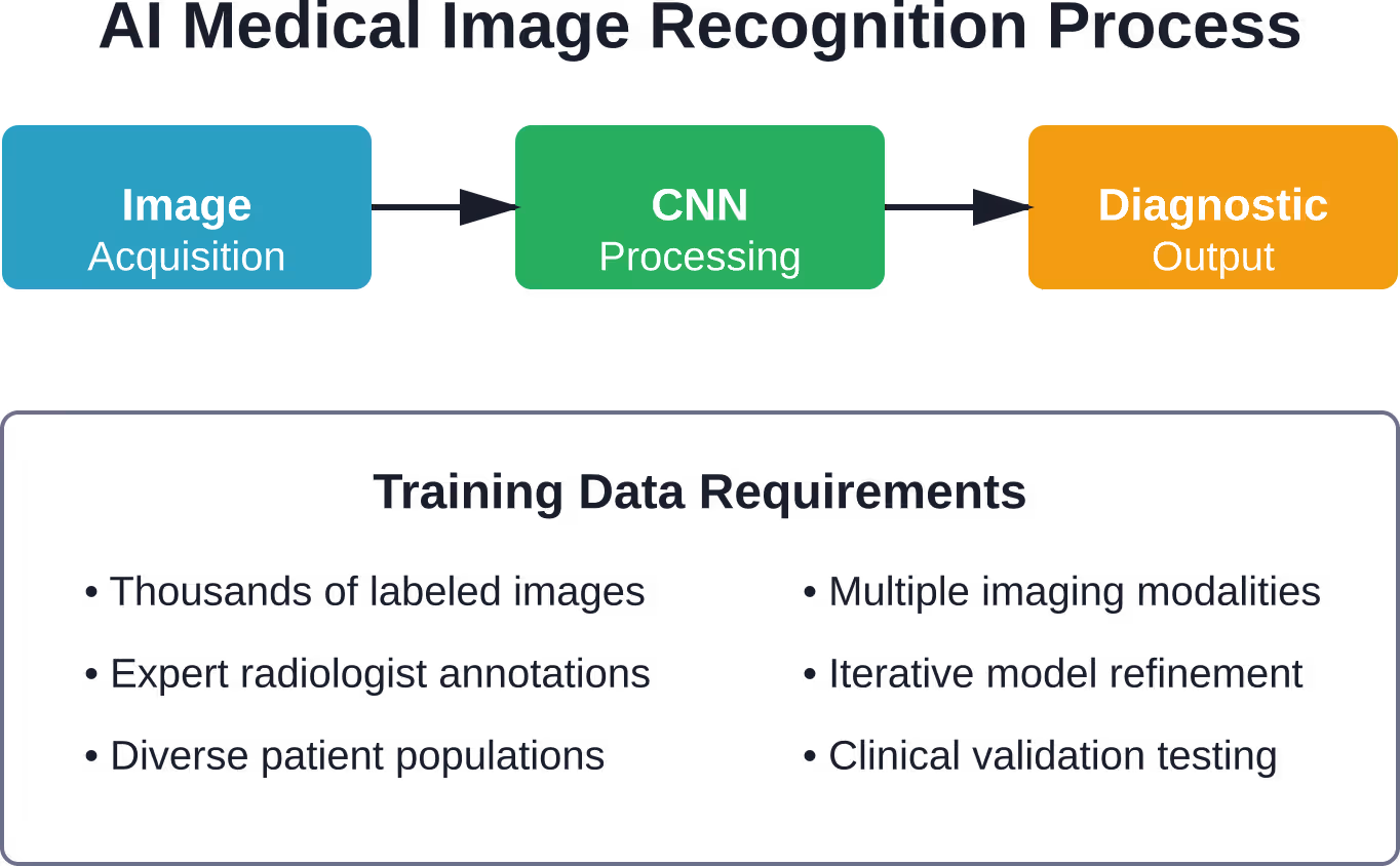
At its core, medical image recognition relies on machine learning models trained on thousands or millions of labeled medical images. The process starts with data collection: hospitals and research institutions compile vast datasets of X-rays, MRIs, CT scans, and other imaging modalities, each annotated by expert radiologists.
These datasets train convolutional neural networks—a type of deep learning architecture specifically designed to process visual information. CNNs automatically learn to identify relevant features in images: edges, textures, shapes, and patterns that correlate with specific medical conditions.
The training process is iterative. The algorithm examines an image, makes a prediction about what it sees, compares that prediction to the expert’s label, and adjusts its internal parameters to improve accuracy. After thousands of iterations across millions of images, the model becomes remarkably skilled at pattern recognition.
Key Technologies Behind Medical Image Recognition
Several technological advances have made AI-driven medical imaging possible. Convolutional neural networks handle automated detection and classification of abnormalities in chest X-rays, lesion detection in dermatology images, and tumor identification in MRI scans.
Transfer learning allows researchers to take models pre-trained on massive general image datasets and fine-tune them for specific medical tasks. This dramatically reduces the amount of labeled medical data required to achieve high accuracy.
Segmentation algorithms go beyond simple classification to outline exact boundaries of tumors, organs, or other structures within medical images. This precision proves invaluable for surgical planning and radiation therapy targeting.

Build Image Recognition Tools With AI Superior
AI Superior develops custom AI software, including computer vision and image processing solutions. Their team can build systems for image analysis, object detection, image segmentation, OCR, face recognition, and contextual image classification.
For medical diagnosis workflows, this can support image-based analysis, scan review, visual classification, or decision-support tools built around clinical data and existing systems.
Need Image Recognition Built Around Your Data?
AI Superior can help with:
- building custom computer vision solutions
- detecting and classifying objects in images
- testing ideas through PoC or MVP development
- integrating AI tools into existing systems
👉 Contact AI Superior to discuss your project.
FDA-Cleared AI Medical Imaging Systems
Regulatory approval is crucial for medical AI systems. The FDA maintains an AI-Enabled Medical Device List that tracks artificial intelligence and machine learning-based devices authorized for marketing in the United States.
Recent clearances demonstrate the breadth of applications. The FDA maintains an AI-Enabled Medical Device List tracking AI-enabled medical devices authorized for marketing in the United States. Multiple advanced systems have received clearance across various specialties including radiology, cardiovascular, and gastroenterology applications. for gastroenterology-urology applications on December 22, 2025.
The FDA AI-Enabled Medical Device List includes cleared systems across multiple specialties including radiology, cardiology, neurology, and gastroenterology. Healthcare providers can reference the FDA’s database for current information on authorized AI-enabled medical devices.
Regulatory Pathways and Standards
Most AI medical imaging systems enter the market through the FDA’s 510(k) clearance pathway, demonstrating substantial equivalence to previously cleared devices. This expedited route allows innovative technologies to reach healthcare providers faster while maintaining safety standards.
The Radiological Society of North America and American College of Radiology have established best practices for AI deployment. RSNA emphasizes that AI tools can play a key role in medical imaging if radiologists trust their design, deploy them with adequate training, and establish clear guidelines regarding clinical accountability.
The ACR Recognized Center for Healthcare-AI program sets guidelines for AI use in imaging interpretation and ensures radiology facilities are using AI safely and effectively. It’s the first national artificial intelligence quality assurance program for radiology facilities designed to recognize adherence to best practices.
Clinical Applications Transforming Patient Care
Image recognition AI has moved beyond research labs into everyday clinical practice. The applications span multiple medical specialties and imaging modalities.
Cancer Detection and Oncology
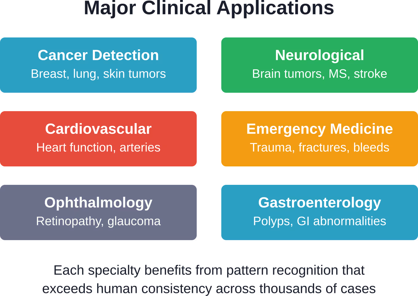
Breast cancer screening has seen particularly impressive results. Deep learning algorithms analyze mammography images to identify suspicious lesions, reducing false negatives and catching early-stage tumors that human readers might miss.
Some studies have shown promising results in detecting referable diabetic retinopathy using AI algorithms with high sensitivity and specificity compared to human graders. Some studies have shown AI algorithms achieving high sensitivity and specificity comparable to or exceeding human graders in detecting referable diabetic retinopathy, significantly reducing the human resources required for screening.
Lung nodule detection in CT scans represents another breakthrough area. AI systems can identify tiny nodules that might indicate early-stage lung cancer, flagging them for radiologist review and enabling earlier intervention.
Neurological and Cardiovascular Imaging
Brain imaging analysis benefits enormously from AI assistance. Algorithms can segment brain tumors, quantify lesion volumes, track disease progression in multiple sclerosis, and identify early signs of neurodegenerative diseases.
Cardiovascular imaging applications include automated measurement of cardiac chamber volumes, ejection fraction calculation, identification of coronary artery calcification, and detection of valve abnormalities. These automated assessments save time and provide consistent, reproducible measurements.
Emergency and Trauma Medicine
Speed matters in emergency departments. AI systems can prioritize critical cases by automatically identifying life-threatening conditions in imaging studies: intracranial hemorrhages, pulmonary embolisms, pneumothorax, and vertebral fractures.
The FDA cleared an Artificial Intelligence Algorithm for the Interpretation of Traumatic injuries, demonstrating regulatory confidence in AI’s ability to assist with time-sensitive diagnoses that can save lives.
Benefits for Healthcare Providers and Patients
The advantages of AI-powered medical image recognition extend throughout the healthcare ecosystem. For radiologists and diagnostic specialists, these systems function as a second set of eyes—catching abnormalities that might be overlooked during long shifts or when reviewing hundreds of studies daily.
Workflow efficiency improves dramatically. Algorithms can pre-screen studies, prioritize urgent cases, perform routine measurements, and generate preliminary reports. This allows specialists to focus their expertise where it matters most: complex cases requiring nuanced clinical judgment.
Consistency is another major benefit. Human readers vary in their interpretations based on experience, fatigue, and individual judgment. AI systems apply the same analytical approach to every image, reducing variability and ensuring standardized assessments.
Expanding Access to Specialized Diagnostics
Geographical disparities in healthcare access represent a persistent challenge. Rural hospitals and underserved communities often lack specialized radiologists, particularly subspecialists in fields like neuroradiology or pediatric imaging.
AI systems can partially bridge this gap. A general radiologist supported by specialized AI tools can provide more accurate diagnoses in areas outside their primary expertise. The technology doesn’t replace specialist training, but it does extend diagnostic capabilities to facilities that might otherwise need to transfer patients or wait days for remote reads.
Telemedicine integration amplifies this effect. Images captured at remote facilities can be analyzed by AI systems in real-time, with concerning findings immediately flagged for expert review regardless of time zones or geographic distance.
Earlier Detection and Better Outcomes
Catching disease early often makes the difference between successful treatment and poor outcomes. AI excels at identifying subtle patterns that indicate early-stage disease—patterns that might not yet be obvious to human observers.
Early cancer detection particularly benefits from this capability. Tumors identified when they’re smaller and haven’t metastasized are dramatically more treatable. The same principle applies to cardiovascular disease, neurodegenerative conditions, and many other progressive illnesses.
Quantitative tracking over time provides another advantage. AI can precisely measure disease progression by comparing sequential imaging studies, detecting changes too subtle for visual comparison. This helps clinicians make informed decisions about treatment adjustments.
Challenges and Limitations
Despite impressive capabilities, medical image recognition AI faces real limitations that healthcare systems must acknowledge.
Data Quality and Bias
AI models are only as good as their training data. If training datasets lack diversity—underrepresenting certain populations, age groups, or disease presentations—the resulting algorithms may perform poorly on underrepresented groups.
This isn’t a hypothetical concern. Community discussions and research publications have documented cases where algorithms trained primarily on one demographic showed reduced accuracy when applied to others. Addressing this requires deliberate effort to compile diverse, representative datasets.
Image quality variability poses another challenge. Different scanners, imaging protocols, and technical settings produce images with varying characteristics. Algorithms trained on images from one institution or equipment manufacturer may struggle when deployed elsewhere.
Interpretability and Clinical Trust
Deep learning models are often described as “black boxes”—they produce predictions without explaining their reasoning. For clinicians accustomed to understanding the basis for diagnostic conclusions, this opacity creates discomfort.
Research into explainable AI aims to address this by generating visualizations showing which image regions most influenced the algorithm’s decision. These attention maps or saliency maps help clinicians understand and verify AI reasoning.
But here’s the reality: even with explanation tools, clinicians need adequate training to work effectively with AI systems. RSNA emphasizes that deployment must include proper education so radiologists can interpret AI outputs appropriately and recognize when to trust or question algorithmic suggestions.
Regulatory and Liability Questions
Who’s responsible when an AI system misses a diagnosis or generates a false positive? Legal frameworks around medical AI liability remain unsettled in many jurisdictions.
Most regulatory guidance treats AI as an assistive tool rather than an autonomous decision-maker. The human physician retains ultimate responsibility for diagnosis and treatment decisions. This provides clarity for liability purposes but also means clinicians can’t simply defer to algorithmic outputs.
Documentation standards need updating too. When AI contributes to a diagnostic decision, how should that be recorded in medical records? What level of detail about the algorithm’s role is appropriate? These operational questions are still being worked out across healthcare systems.
| Challenge Area | Impact | Current Solutions |
|---|---|---|
| Data Bias | Reduced accuracy for underrepresented groups | Diverse training datasets, demographic testing |
| Model Interpretability | Difficulty understanding AI reasoning | Attention maps, explainable AI methods |
| Regulatory Compliance | Complex approval pathways | FDA 510(k), quality assurance programs |
| Clinical Integration | Workflow disruption, training requirements | PACS integration, radiologist education |
| Equipment Variability | Inconsistent performance across scanners | Multi-site training, protocol standardization |
Implementation Considerations for Healthcare Facilities
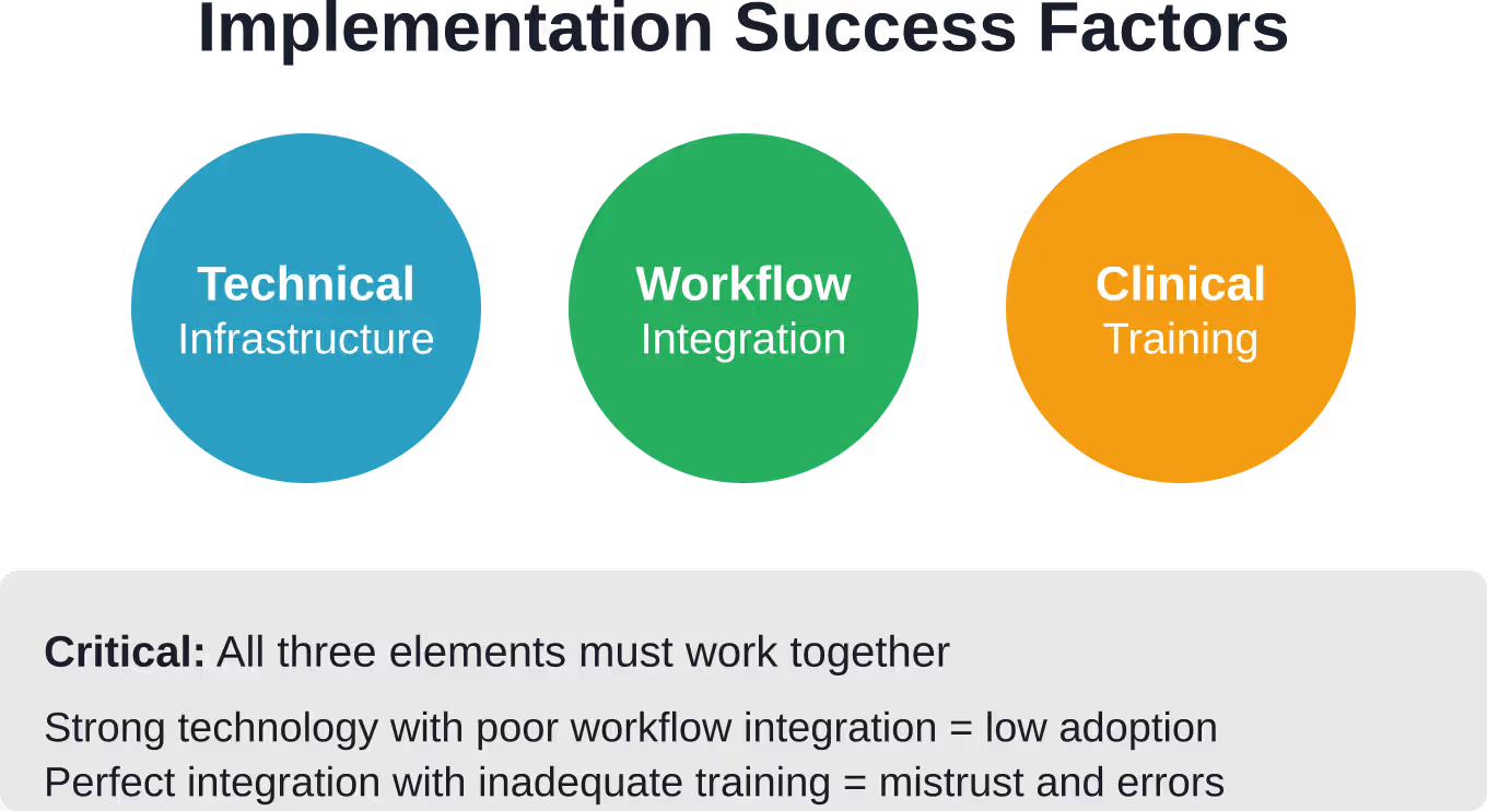
Adopting AI image recognition requires more than purchasing software. Successful implementation demands careful planning across technical, clinical, and operational dimensions.
Technical Infrastructure
AI systems need computational resources—either on-premises GPU servers or cloud computing capacity. Integration with existing Picture Archiving and Communication Systems is essential so algorithms can access images and deliver results within radiologists’ normal workflow.
Network bandwidth matters too, especially for cloud-based solutions. High-resolution medical images are large files. Uploading studies for AI analysis and receiving results must happen quickly enough to support clinical decision-making timelines.
Data security and privacy protections require particular attention. Patient imaging data is protected health information subject to HIPAA regulations in the United States and similar privacy laws elsewhere. Any AI implementation must include appropriate safeguards.
Clinical Workflow Integration
The best AI system fails if clinicians won’t use it. Workflow integration determines adoption success. Algorithms should present results within existing interfaces—embedded in PACS workstations or integrated into radiology information systems rather than requiring separate applications.
Result presentation format matters. Overwhelming radiologists with excessive information or alerts leads to alarm fatigue and ignored recommendations. Effective systems present findings clearly, prioritize genuinely concerning cases, and minimize false positives.
Staff training can’t be an afterthought. Radiologists, technologists, and referring physicians all need education about AI capabilities, limitations, and appropriate use. This includes understanding when to trust AI outputs and when to apply additional scrutiny.
Validation and Ongoing Monitoring
Before clinical deployment, healthcare facilities should validate AI system performance on their own patient population and imaging equipment. Performance characteristics may differ from vendor-reported metrics based on different demographics or technical factors.
Ongoing monitoring after deployment is equally important. AI systems can degrade over time if patient populations shift, equipment is upgraded, or imaging protocols change. Periodic audits comparing AI outputs against expert human reads help identify performance drift.
Quality assurance programs like the ACR’s ARCH-AI provide frameworks for systematic validation and monitoring, helping facilities maintain high standards as they incorporate AI into routine practice.
The Future of AI in Medical Imaging
Current AI systems focus primarily on detection and classification—identifying what’s in an image. The next generation will move toward prediction and decision support.
Multimodal Integration
Future systems will combine imaging data with electronic health records, genomic information, and other data sources. This holistic approach enables more sophisticated analysis: not just identifying a tumor, but predicting its likely behavior based on imaging characteristics correlated with molecular profiles.
Natural language processing will extract relevant clinical context from radiology reports and medical notes, incorporating that information into image interpretation. This mimics how expert radiologists work—considering clinical history alongside visual findings.
Real-Time Surgical Guidance
AI image recognition is beginning to enter operating rooms. Real-time analysis of surgical video can help identify anatomical structures, detect complications, and guide instrument placement. Augmented reality systems overlay AI-generated guidance onto the surgeon’s view.
These applications require extreme reliability and near-instantaneous processing. The technical challenges are significant, but early results show promise for improving surgical precision and reducing complications.
Personalized Medicine
Imaging biomarkers identified through AI analysis may enable truly personalized treatment selection. Rather than treating all patients with similar diagnoses the same way, clinicians could tailor interventions based on imaging features that predict treatment response.
This approach is already showing results in oncology, where tumor imaging characteristics help predict which patients will benefit from specific chemotherapy regimens or immunotherapy approaches.
Frequently Asked Questions
How accurate is AI for medical diagnosis compared to human radiologists?
Accuracy depends heavily on the specific task and algorithm. For some narrowly defined applications—like detecting diabetic retinopathy or certain types of lung nodules—AI systems have demonstrated accuracy equal to or exceeding experienced specialists. However, radiologists still outperform AI on complex cases requiring integration of multiple findings or clinical context. The most effective approach combines AI detection with human expertise and judgment.
Will AI replace radiologists and other diagnostic specialists?
No. AI systems function as assistive tools that enhance radiologist capabilities rather than replacing them. Radiologists provide clinical context, integrate findings across multiple studies, communicate with referring physicians, and make nuanced judgments that AI cannot replicate. The technology shifts radiologist work toward higher-value activities: complex case interpretation, procedure guidance, and clinical consultation.
Are AI medical imaging systems regulated and tested for safety?
Yes. In the United States, the FDA regulates AI-enabled medical devices through its standard clearance and approval pathways. Most AI imaging systems receive 510(k) clearance after demonstrating substantial equivalence to previously cleared devices. The FDA maintains an AI-Enabled Medical Device List tracking authorized systems. Similar regulatory oversight exists in Europe, Canada, and other jurisdictions. Healthcare facilities can also participate in quality assurance programs like ACR’s ARCH-AI to ensure best practices.
What medical specialties benefit most from AI image recognition?
Radiology sees the highest volume of AI applications, including detection of fractures, tumors, and vascular abnormalities across X-ray, CT, and MRI. Ophthalmology benefits significantly from automated retinal image analysis for diabetic retinopathy and glaucoma. Pathology uses AI for analyzing tissue slides. Cardiology applies AI to echocardiograms and cardiac MRI. Gastroenterology employs AI during colonoscopy to detect polyps. Essentially, any specialty that relies heavily on medical imaging can benefit.
How much does AI medical imaging software cost?
Pricing varies widely based on application, deployment model, and volume. Some vendors offer per-study pricing ranging from a few dollars to over twenty dollars per scan. Others use subscription models with annual fees. Enterprise licenses for healthcare systems can reach hundreds of thousands of dollars annually. For current pricing specific to particular systems, check vendor websites directly as costs change frequently.
Can AI detect diseases earlier than traditional methods?
In some cases, yes. AI excels at identifying subtle patterns that may indicate early-stage disease before it becomes obvious to human observers. This has proven particularly valuable for early cancer detection, where small tumors or precancerous changes might be missed visually. However, earlier detection only improves outcomes when effective treatments exist for early-stage disease. AI detection capabilities must be validated through clinical studies demonstrating actual patient benefit.
What are the main limitations of current AI diagnostic systems?
Key limitations include dependence on training data quality, potential bias if datasets lack diversity, difficulty generalizing across different scanner types or imaging protocols, limited interpretability of algorithm reasoning, inability to incorporate clinical context as effectively as human specialists, and challenges with rare conditions not well-represented in training data. These systems also generate false positives and false negatives, requiring human oversight.
Moving Forward with Medical Image Recognition AI
Image recognition for medical diagnosis represents one of healthcare’s most promising technology frontiers. The combination of deep learning algorithms and massive imaging datasets has produced systems that genuinely assist clinicians in detecting disease earlier and more consistently.
But the technology isn’t magic. It requires careful implementation, ongoing validation, proper training, and realistic expectations about capabilities and limitations.
Healthcare facilities exploring AI adoption should start with clear use cases addressing specific clinical needs—not technology for technology’s sake. Successful deployment demands collaboration between IT teams, radiologists, administrators, and vendors. Quality assurance frameworks ensure systems continue performing as expected over time.
The hundreds of FDA-cleared AI imaging devices now available demonstrate regulatory confidence and clinical demand. Recent clearances through December 2025 span radiology, cardiology, gastroenterology, and other specialties, reflecting AI’s expanding role across medical imaging.
For healthcare providers, staying informed about AI capabilities and limitations is essential. For patients, understanding that AI assists rather than replaces physician expertise provides appropriate context. The human-AI partnership in medical diagnosis is just beginning, with tremendous potential to improve care quality and accessibility in the years ahead.
 English
  German
  Arabic
  French
  Dutch
Dutch  Spanish