The challenge
An ophthalmology centre needed to estimate the volume of fat and muscle in the human eye accurately. This information is essential for monitoring eye health, evaluating the effectiveness of interventions and carrying out comparative analyses.
Estimating these volumes relied on manual measurement, which was time-consuming and prone to error. The centre wanted an automated, reliable way to handle this part of eye care.
Our solution
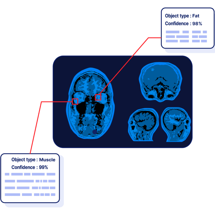
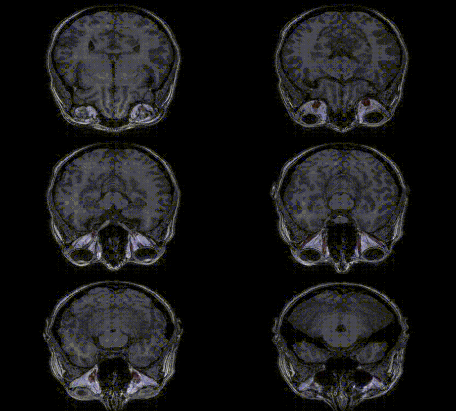
We focused on MRI orbit scans and developed a deep learning model that segments fat and muscle tissue in each slice of the scan. Summing the segmented tissue across slices gives a precise volume estimate for these anatomical structures of the eye.
The model handles the coronal, sagittal and axial views of the eye, so the whole orbital region is covered.
- Slice-by-slice segmentation of fat and muscle tissue in MRI orbit scans
- Volume estimation of fat and muscle from the segmented slices
- Support for coronal, sagittal and axial views
- Before-and-after volume comparison following an intervention
The outcome
Automating the segmentation of fat and muscle tissue gives ophthalmologists precise volume estimates without manual measurement, supporting more comprehensive assessments.
Because the model enables before-and-after comparisons, clinicians can quantify changes in fat and muscle volume and assess the impact of surgical procedures, therapeutic interventions or disease progression on the ocular structures. The project complements our Eye Metrics work in ophthalmology.